Correction Surgery for a Blunt and Wide Nose_Rhinoplasty Korea
- noselab
- 2024년 10월 25일
- 3분 분량
최종 수정일: 4월 7일
Oblique View:
Hello, this is Dr. Cha-Young Kang, chief surgeon at Nose Lab Clinic.
Today, I’d like to share a case involving a patient who experienced nasal bridge pain, headaches, and discomfort from a loose implant. This patient had undergone an L-shaped silicone implant rhinoplasty 20 years ago and had recently been struggling with chronic rhinitis, which required long-term medication.
1. Pre-Surgery Consultation
Main Symptoms:
Pain around the nasal bridge, especially when wearing glasses.
Noticeable movement of the silicone implant.
Occasional pain in the nasal dorsum (bridge).
Severe rhinitis.
Pre-Surgery Analysis:

Frontal View:
Swollen, wide nasal bridge.
Blunt and droopy nasal tip.
The nasal alae (sides) were flared and droopy.
The philtrum appeared elongated.

Oblique View:
The nasal tip appeared blunt and droopy.
Signs of protrusion of the upper lip.

Side View:
The nasal tip was compressed and blunt.
Columellar retraction and protrusion of the upper lip were noticeable.
Observed signs of a weak chin (microgenia).
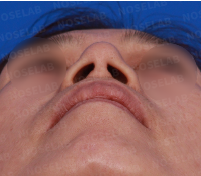
Nostril View:
Slight asymmetry in the nostrils.
Nostrils appeared flattened and wide.
2. Surgical Plan
To address the patient's concerns, we designed the following surgical plan:
Remove the existing silicone implant.
Excise excess tissue surrounding the implant.
Insert a new, thinner, and broader implant.
Elevate the nasal tip and adjust the nasolabial angle.
Perform septal correction and extension graft.
3. Surgical Procedures
1. Removal of Existing Implant:
Removed a hard silicone implant measuring 8mm in width and 3–4mm in thickness.
Excised the surrounding granulation tissue and thoroughly irrigated the area with saline solution.
2. Nasal Tip Reconstruction:
Removed scar tissue around the alar cartilage.
Utilized autologous rib cartilage for septal extension and nasal tip correction.
3. Nasolabial Angle Adjustment:
Dropped the columella and adjusted the nasolabial angle to create a more balanced profile, with an expected improvement in the appearance of the protruding upper lip.
4. Placement of New Implant:
Inserted a new, minimally elevated, and broader silicone implant.
Placed the implant under the periosteum for enhanced stability.
5. Septal Correction:
Corrected only the deviated portion of the septum to preserve as much cartilage as possible.
4. Pre- and Post-Surgery Comparison

Before (Left) / After (Right)
Frontal View:
The previously wide and swollen nasal bridge has slimmed down, and the swelling has reduced.
The blunt nasal tip has been elevated for a more refined appearance.
The flared alar base has been improved.

Before (Left) / After (Right)
Oblique View:
The droopy nasal tip has been corrected, achieving a more balanced height.
The protruding appearance of the upper lip has been reduced.

Before (Left) / After (Right)
Side View:
The nasal tip has been naturally elevated, eliminating the blunt appearance.
The columellar retraction has been corrected, and the nasolabial angle has been adjusted to a more natural position.
The overall balance of the nose and face has improved, reducing the prominence of the upper lip.

Nostril View:
Nostril asymmetry has been corrected.
The previously flattened nostrils now have a more rounded, natural shape.
5. Immediate Post-Op Photos

Photos Immediately After Surgery: Frontal, Side, and Oblique Side Views

Photos Immediately After Surgery: Nostril View
6. Conclusion
This case involved addressing complications from an outdated rhinoplasty procedure (pain, implant movement) while providing cosmetic improvement. The surgery included implant replacement, nasal tip reconstruction using autologous rib cartilage, and nasolabial angle correction to achieve both functional and aesthetic goals.
For this patient, it was crucial to eliminate the chronic foreign body response, which likely contributed to neuralgia-like pain. We focused on removing the source of irritation and reducing pressure in the affected areas. Over the next six months, we will closely monitor the patient’s recovery, and additional adjustments will be made if necessary.
7. Final Thoughts
Post-rhinoplasty patients may occasionally experience pressure or pain around the nasal bridge, often occurring within the first six months of surgery. In many cases, these symptoms improve over time as scar tissue subsides. For some patients, however, the thickness of the implant may exceed what the nasal bridge can comfortably support, in which case a reduction in implant height can resolve the issue.
When nasal bridge pain occurs years or even decades after surgery, it could indicate a chronic foreign body reaction or an acute or chronic infection. In such cases, surgical intervention to assess the nasal bridge, reposition the implant, and clean the area is often sufficient to resolve the problem.
If you are considering rhinoplasty or experiencing discomfort from a previous surgery, consult with a specialist to determine the root cause and the most appropriate solution.
At Nose Lab Clinic, we prioritize the safety and satisfaction of our patients, striving for the best possible outcomes
This has been Dr. Cha-Young Kang, chief surgeon at Nose Lab Clinic.
Thank you.


댓글