Case at a glance
- Surgical plan
- Graft & material details
- Photo timing
These details are excerpts from the original article and captions. Follow a detail to read it in context.
Hello, this is Dr. Cha-Young Kang, Chief Director at NoseLab Plastic Surgery Clinic.
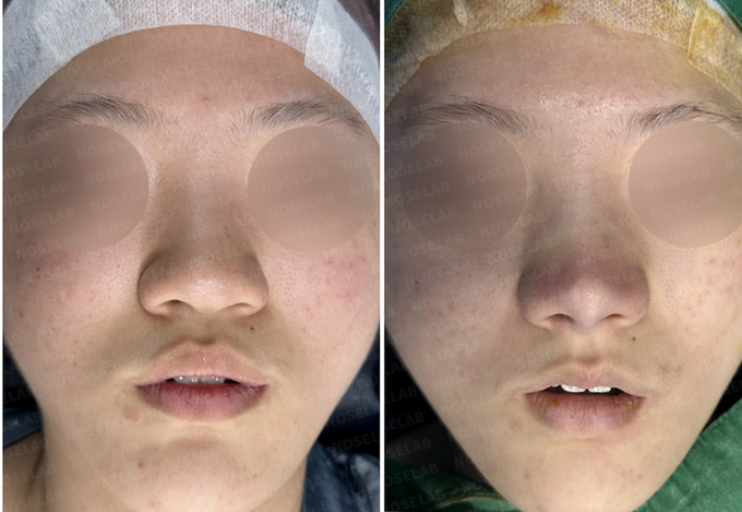
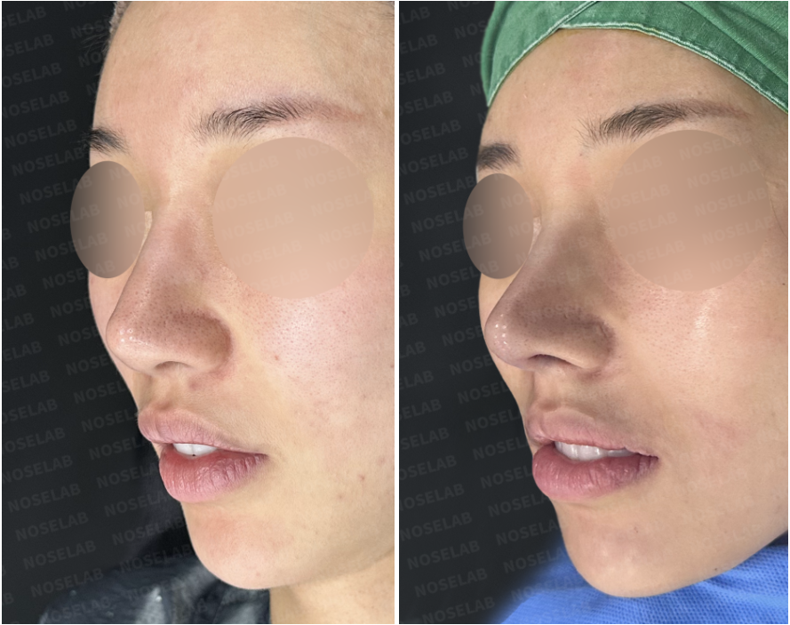
Today, I am sharing a Visible Cartilage Revision Rhinoplasty Korea case involving visible nasal tip cartilage, a wide nasal framework, and functional nasal obstruction years after primary rhinoplasty. This revision focused on restoring structural balance, reinforcing thin skin, and achieving long-term functional stability.

Surgical Background of Visible Cartilage Revision Rhinoplasty Korea
The patient underwent comprehensive rhinoplasty approximately 7 years ago, including silicone implant placement, ear cartilage grafting, septal cartilage manipulation, and alar base reduction. Over time, thin nasal tip skin and pointed cartilage grafts led to visible cartilage under light exposure, accompanied by a wide nasal appearance and nasal obstruction caused by septal deviation and turbinate hypertrophy.

Surgical Plan
Autologous Dermis Grafting
The patient’s own dermis was grafted over the nasal tip to thicken the skin layer, eliminating cartilage visibility while preserving natural texture. Autologous dermis offers long-term volume stability without foreign body reactions.
Cartilage Reshaping and Redistribution
Existing cartilage grafts were reshaped and widened to reduce pressure against the skin, preventing recurrence while maintaining a refined nasal tip.
Tip Refinement with Rib Cartilage
Autologous rib cartilage was used to sculpt a sharp yet stable nasal tip and support a near-straight dorsal line.
Functional Corrections
Lateral osteotomy corrected wide and deviated nasal bones, while septoplasty and radiofrequency turbinate reduction restored nasal airflow.
Surgical Results
Complete resolution of visible cartilage
Slimmer, symmetrical nasal framework
Refined, sharp nasal tip with natural skin contour
Significant improvement in nasal breathing
Stable results without recurrence



Expert Commentary
Visible cartilage is most commonly caused by thin skin combined with sharp or narrowly placed grafts. In revision cases, masking the problem is ineffective. Reinforcing the skin with autologous dermis and reshaping the cartilage structure is essential for durable results.

Read the author’s background and the original case context. About these articles