Closed Rhinoplasty Korea for Contracted Nose Reconstruction and Functional Recovery
- Dr. Chayoung Kang
- 2024년 7월 29일
- 2분 분량
최종 수정일: 6일 전
This case demonstrates closed rhinoplasty Korea for severe nasal contracture following infection and implant removal. Structural reconstruction using rib cartilage and functional correction restored nasal symmetry, improved airway function, and achieved long-term stability.
Author: Dr. Chayoung Kang, Director, NoseLab Rhinoplasty Clinic
This patient developed severe nasal contracture and airway obstruction following infection after previous rib cartilage rhinoplasty.
The implant had to be removed within two weeks due to inflammation, leaving the nasal structure severely compromised.
In this closed rhinoplasty Korea case, the goal was to restore structural integrity and improve nasal function through staged reconstruction.
Key concerns included:
Severe nasal contracture
Loss of septal support
Nostril asymmetry
Hardened nasal tip tissue
Nasal obstruction and mouth breathing
Internal adhesions
📩 International Consultation


✅ Surgical Background – Closed Rhinoplasty Korea
Closed rhinoplasty Korea enables precise reconstruction of damaged nasal structures without external incision.
In contracture cases, both structural loss and scar contraction must be addressed simultaneously.
✅ Structural Problems Identified
Preoperative analysis revealed:
Severe contracture with hardened tissue
Loss of septal cartilage
Alar cartilage deficiency
Nostril asymmetry
Internal mucosal adhesion
Functional airway obstruction

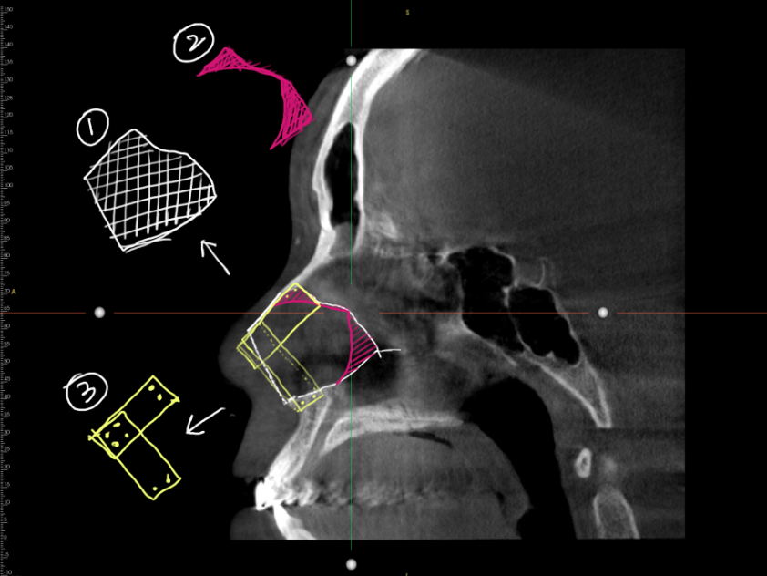
✅ Surgical Plan
1. Preoperative Tissue Preparation
Stem cell therapy was performed to improve tissue quality.
2. Osteotomy
Correction of nasal bone asymmetry.
3. Septal Reconstruction
Rebuilt using rib cartilage.
4. Alar Reconstruction
Restored nostril symmetry.
5. Columella Adjustment
Normalized nasal base balance.
6. Adhesion Release
Improved airway function.


✅ Surgical Results
Frontal View
Improved symmetry and balanced structure

Lateral View
Restored projection and natural angle

Oblique View
Reduced contracture and improved contour

Nasal Base View
Improved nostril symmetry

Functional Outcome
Improved breathing
Reduced obstruction
Stable airway
✅ Surgeon’s Commentary
Nasal contracture caused by infection is one of the most complex conditions in rhinoplasty.
The main issue is not only structural loss but also scar contraction and tissue damage.
Rib cartilage reconstruction is essential to rebuild stable nasal support in such cases.
Preoperative tissue preparation significantly improves surgical outcomes.
Closed rhinoplasty Korea allows precise correction while minimizing additional damage.
If you are experiencing similar complications, a structural approach is essential.
📩 International Consultation
FAQ
Q1. What causes nasal contracture after rhinoplasty?
Nasal contracture usually results from postoperative infection or severe inflammation that leads to scar formation and cartilage loss.
Q2. Why is rib cartilage used in reconstructive cases?
Autologous rib cartilage provides sufficient strength and volume to rebuild severely damaged nasal structures and maintain long-term stability.
Q3. Can nasal breathing improve after contracture surgery?
Yes. Releasing adhesions and correcting septal deviation can significantly improve nasal airflow and reduce mouth breathing.

Messenger(WhatsApp) : +82 1057360302
Home page : www.noselab.co.kr
Instagram : noselab_global
YouTube : Noselab
Email : noselab@naver.com


댓글