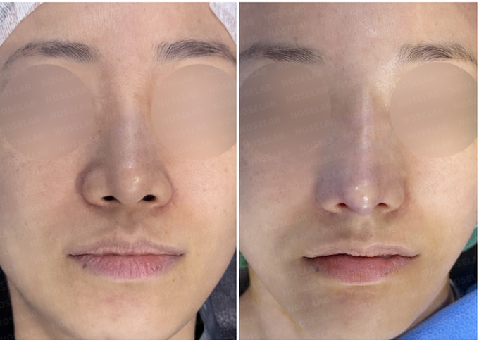
Case at a glance
- Concerns & goals
- Previous treatment
- Surgical plan
- Graft & material details
- Photo timing
These details are excerpts from the original article and captions. Follow a detail to read it in context.
This case reviews nasal tip and bridge translucency correction in a patient who developed thin nasal skin, visible nasal tip cartilage, bridge translucency, and a chalky nasal appearance after long-term L-shaped silicone implant rhinoplasty. Surgery focused on implant removal, autologous rib cartilage nasal tip reconstruction, dermal grafting, skin reinforcement, implant replacement, and dorsal contour correction.
Author: Dr. Cha-Young Kang
Clinic: NoseLab Clinic
Published: 2025
Last Updated: 2026
Introduction
Hello, this is Dr. Cha-Young Kang of NoseLab Clinic.
This case involves nasal tip and bridge translucency correction in a patient who had undergone L-shaped silicone implant rhinoplasty 18 years earlier. Over time, the patient developed thinning of the nasal skin, visible cartilage outline at the nasal tip, bridge translucency, and a pale chalky appearance along the nose.
The surgery was performed using a closed rhinoplasty / endonasal approach. The goal was to reduce implant-related pressure, reinforce thin skin, reconstruct the nasal tip with autologous rib cartilage, and improve the nasal bridge contour using dermal grafting and implant replacement.
Case Background of Nasal Tip and Bridge Translucency Correction
The patient had a long surgical history involving filler injection, L-shaped silicone implant rhinoplasty, ear cartilage grafting, filler removal, and AlloDerm grafting.
Surgical History
The surgical history included:
19 years ago: Nasal filler injection
18 years ago: L-shaped silicone implant, ear cartilage graft, and filler removal
6 months later: AlloDerm graft due to nasal translucency
Despite previous soft tissue reinforcement, the patient continued to have visible nasal tip and bridge translucency over time.
Because the case involved both thin nasal skin and implant-related contour problems, the revision plan needed to address the skin layer, nasal tip support, and dorsal implant contour together.
Initial Concerns
Preoperative design consultation photos were reviewed from the frontal, side, oblique, and nostril views.
Main Concerns
The main concerns included:
Nasal tip cartilage visible through the skin
Nasal bridge translucency caused by a narrow and elevated silicone implant
Thin nasal skin
Rigid nasal soft tissue
Pale chalky appearance along the nose
Harsh overall nasal impression
Long-term implant-related pressure
These findings suggested that simple implant exchange would not be enough. The thin skin and structural support needed to be corrected together.

Preoperative Design Assessment
Frontal View
The frontal view showed a sharp and pale appearance along the nasal bridge and tip area, suggesting thin skin coverage over the underlying implant and cartilage.
Side View
The side view showed an elevated, narrow nasal bridge line and implant-related tension along the dorsum and nasal tip.
Oblique View
The oblique view showed visible contour irregularity and translucency along the nasal bridge and tip transition.
Nostril View
The nostril view was used to assess nasal tip support, skin tension, and lower nasal balance.
The surgical plan was designed to reduce skin tension, improve soft tissue coverage, and rebuild nasal tip support.

Surgical Plan for Translucency Correction
The surgical plan focused on reducing implant-related pressure, reinforcing the thin nasal skin, reconstructing the nasal tip, and improving the dorsal contour.
Skin Tension Adjustment
The nasal tip was lowered to reduce excessive pressure on the thin skin.
Reducing skin tension was important because long-term pressure from an L-shaped silicone implant can make the nasal tip skin thinner and more vulnerable.
Dermal Grafting for Thin Skin Reinforcement
A dermal graft was applied to reinforce the thinned nasal skin.
This helped provide additional soft tissue coverage over areas where cartilage or implant edges were visible through the skin.
Nasal Tip Reconstruction with Autologous Rib Cartilage
Autologous rib cartilage was used to reconstruct the nasal tip structure.
The cartilage framework was designed to support the tip while distributing contact pressure over a wider area.
This was important because narrow contact points can make light reflection and contour visibility more noticeable under thin skin.
Implant Removal
The previous silicone implant was removed.
This allowed reassessment of the nasal bridge, skin thickness, and implant-related contour changes.

Bridge Refinement and Implant Replacement
The previous rigid and narrow implant was replaced with a broader and softer dorsal material.
The goal was to reduce visible edge contrast and create a smoother transition along the nasal bridge.
Tissue Grafting for Dorsal Coverage
Additional tissue grafting was used to cover and support the nasal bridge.
This helped improve soft tissue coverage and reduce the chalky appearance caused by thin skin and implant show-through.
Post-Surgical Results of Tip and Bridge Correction

Nasal Tip
At the nasal tip, translucency and visible cartilage outline were reduced after rib cartilage reconstruction and dermal grafting.
The nasal tip width and height appeared more balanced after pressure redistribution and soft tissue reinforcement.

Nasal Bridge
At the nasal bridge, the contour appeared smoother after implant removal, implant replacement, and additional tissue coverage.
The chalky appearance along the bridge was reduced after improving soft tissue coverage and dorsal contour.

Overall Appearance
The transition between the nasal tip and bridge appeared smoother after combined structural and soft tissue correction.
The overall nasal appearance became softer after reducing visible implant-related edges and reinforcing the thinned skin.

Structural Outcome
The nasal tip was supported with autologous rib cartilage, and the thin skin was reinforced with dermal grafting.
Because this case involved long-term implant use and thin nasal skin, the outcome was planned within the limits of skin thickness, tissue rigidity, implant history, and healing response.
Surgeon’s Commentary
This case shows a long-term complication pattern that can occur after L-shaped silicone implant rhinoplasty. When an implant extends toward the nasal tip or creates narrow pressure points, the skin may become thin over time.
Thin skin can make cartilage edges, implant borders, or dorsal contour irregularities more visible. This may appear as nasal tip translucency, bridge translucency, or a chalky nasal appearance.
In this case, the surgical plan required both structural reconstruction and soft tissue reinforcement. The previous implant was removed, and the nasal tip was reconstructed using autologous rib cartilage.
Dermal grafting was used to improve soft tissue coverage over the thin skin. A broader dorsal material and additional tissue grafting were used to reduce visible edge contrast along the nasal bridge.
This case shows that nasal tip and bridge translucency correction should be planned as a combined implant, cartilage, skin, and soft tissue procedure rather than simple implant replacement alone.
FAQ
Why does nasal tip or bridge translucency occur after rhinoplasty?
Nasal translucency can occur when the skin becomes thin or when cartilage or implant edges are close to the skin surface. Long-term implant pressure, previous surgery, and skin thinning can make this more visible.
Can L-shaped silicone implants cause thin nasal tip skin?
An L-shaped silicone implant may place pressure on the nasal tip if it extends too far downward. Over time, this pressure can contribute to thinning of the nasal tip skin in some patients.
How is nasal tip translucency corrected?
Correction may include reducing skin tension, reconstructing the nasal tip, reinforcing thin skin with dermal grafting, and adjusting the contact area between cartilage and skin.
Why is rib cartilage used in translucency correction?
Rib cartilage may be used when stronger structural support is needed for nasal tip reconstruction. It can help create a broader support framework and reduce sharp contact points under thin skin.
Why is dermal grafting used for thin nasal skin?
Dermal grafting may help reinforce thin skin and provide additional soft tissue coverage. It can be used when cartilage edges, implant outlines, or contour irregularities are visible through the skin.
International Consultation
For international patients, a photo-based consultation may help clarify whether nasal tip and bridge translucency correction, implant visibility revision rhinoplasty, rib cartilage reconstruction, dermal grafting, or implant replacement may be needed.
Read the author’s background and the original case context. About these articles