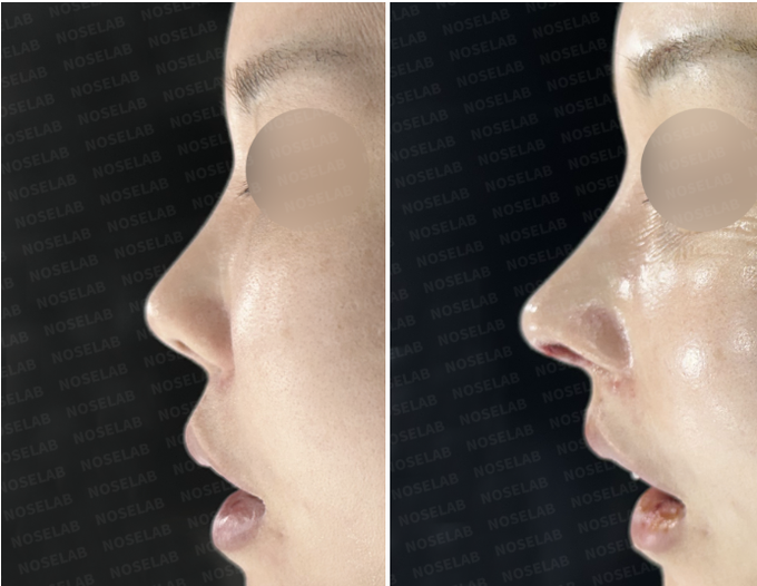
Case at a glance
- Concerns & goals
- Previous treatment
- Approximately 20 years earlier, the patient underwent silicone implant placement, septal surgery, and alar base reduction. Right-sided nasal obstruction became more severe afterward.
- Approximately 10 years earlier, the patient underwent another operation for a deviated septum at a different clinic, but difficulty breathing through the right side persisted.
- Surgical plan
- Septal perforation repair: The previous two operations had left the septal cartilage extensively harvested and excessively scored, compromising its structural stability.
- Septal reconstruction with the patient's own rib cartilage: We harvested autologous rib cartilage to rebuild and reinforce the damaged septum.
- Silicone implant replacement and nasal osteotomies: The existing implant was removed and replaced with a new silicone implant carefully carved to fit the nasal bones.
- Graft & material details
- Septal perforation repair: The previous two operations had left the septal cartilage extensively harvested and excessively scored, compromising its structural stability.
- Septal reconstruction with the patient's own rib cartilage: We harvested autologous rib cartilage to rebuild and reinforce the damaged septum.
- Photo timing
These details are excerpts from the original article and captions. Follow a detail to read it in context.
Hello, I am Dr. Peter C. Kang, Medical Director of NOSELAB Clinic. Today, I would like to share a closed revision rhinoplasty case involving longstanding nasal obstruction, damage to the nasal septum, and a visibly crooked nose.


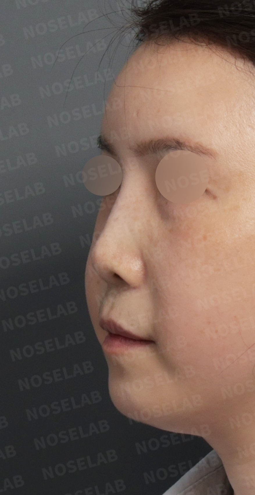
Surgical History
Approximately 20 years earlier, the patient underwent silicone implant placement, septal surgery, and alar base reduction. Right-sided nasal obstruction became more severe afterward.
Approximately 10 years earlier, the patient underwent another operation for a deviated septum at a different clinic, but difficulty breathing through the right side persisted.
Nasal pain began about a year before this consultation, prompting the patient to seek treatment for the underlying problems.

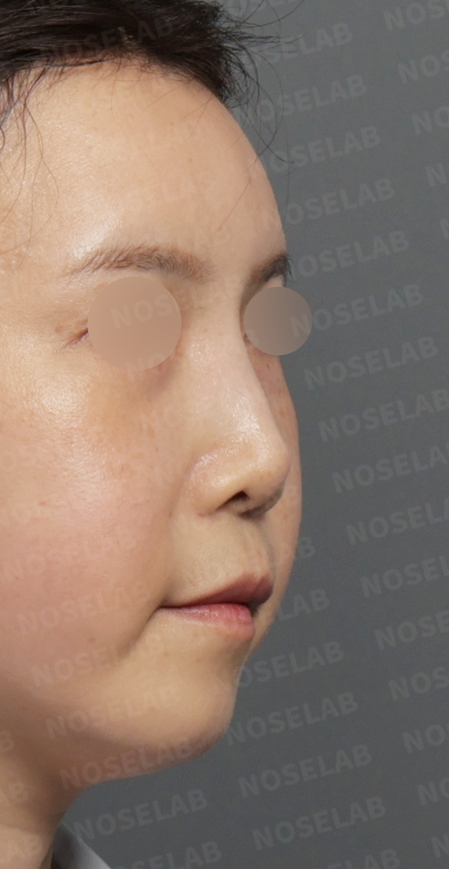
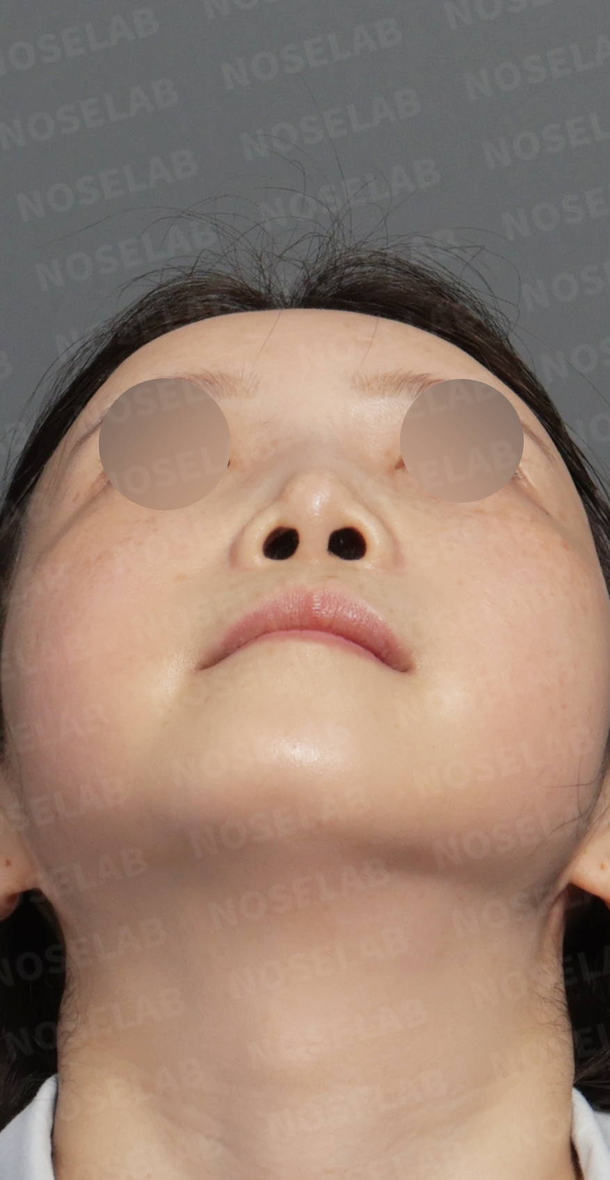
The Patient's Goals
Straighten the crooked nose.
Reduce the appearance of a protruding mouth.
Reduce nostril show from the front.
Improve the appearance of a short nose.
Achieve a natural, straight nasal contour.
Surgical Plan and Procedure
Septal perforation repair: The previous two operations had left the septal cartilage extensively harvested and excessively scored, compromising its structural stability. We also identified a 7 × 5 mm septal perforation, a hole in the nasal septum. This was repaired using perichondrium, the tissue covering cartilage, harvested from the ear.
Septal reconstruction with the patient's own rib cartilage: We harvested autologous rib cartilage to rebuild and reinforce the damaged septum. Its strength made it suitable for establishing durable nasal support and helping the reconstructed framework maintain its shape.
Silicone implant replacement and nasal osteotomies: The existing implant was removed and replaced with a new silicone implant carefully carved to fit the nasal bones. Lateral osteotomies were also performed to correct the crooked, broad appearance of the nose.
Right nasal valve reconstruction and correction of the deviated septum: We reconstructed the narrowed right nasal valve and corrected the septal deviation to address the structural causes of nasal obstruction.
Nasolabial angle correction: The nasal tip and the angle between the nose and upper lip were adjusted to improve the low, short-looking tip and excessive frontal nostril show.
Surgical Results


Frontal view
The crooked nasal contour became straighter and more balanced.
Excessive nostril show was visibly reduced.
Osteotomies refined the previously broad nose into a more defined contour.


Profile view
The short, low-looking tip gained natural projection, creating a more refined profile.
Adjusting the nasolabial angle softened the appearance of a protruding mouth.


Functional changes
Reconstruction of the right nasal valve and correction of the septal deviation improved the longstanding nasal obstruction.
Repairing the septal perforation restored the integrity of the internal nasal framework.
Surgeon's Perspective
This was a complex reconstructive operation that addressed substantial internal damage from previous surgery as well as the external appearance of the nose.
In this patient, the septal perforation required repair because the internal framework was already compromised and could weaken further. We closed the defect using perichondrium harvested from the ear and reconstructed the damaged septal cartilage with the patient's own rib cartilage. Both septal deviation and nasal valve narrowing contributed to the breathing difficulty, so both needed to be addressed. A precise surgical plan that considered appearance and breathing together was central to this case.
Thank you for reading. I am Dr. Peter C. Kang, Medical Director of NOSELAB Clinic.
Information About These Photographs and Surgical Risks
These before-and-after photographs show an individual patient's results. Discuss your expected outcome with your doctor.
All surgery carries a risk of complications, including the following:
Infection: Disinfection and injectable or oral antibiotics are used to help prevent infection. In rare cases (less than 0.5%), another operation may be required.
Bleeding: Nasal packing is used for one to two days to help minimize bleeding. Bleeding may occasionally occur after surgery for nasal inflammation or obstruction.
Allergic reactions.
Residual asymmetry: Facial asymmetry and the condition of the nose may prevent complete correction, as explained during consultation.
Insufficient cosmetic or functional improvement.
Other complications, including excessive scar formation and skin necrosis.


Read the author’s background and the original case context. About these articles