Safe Silicone Exposure Rhinoplasty and Nasal Inflammation Correction
- noselab
- 2024년 10월 22일
- 2분 분량
최종 수정일: 2025년 9월 29일
Hello, this is Dr. Cha-Young Kang, head surgeon at Nose Lab Clinic.
Today, I would like to share a successful case of silicone exposure rhinoplasty where we treated a patient suffering from implant extrusion and nasal inflammation. The patient had undergone rhinoplasty at another clinic 12 years ago with silicone and ear cartilage. Over the past five months, redness and swelling appeared, indicating infection and exposure of the implant.
Pre-Surgery Analysis of Silicone Exposure Rhinoplasty Cases

Frontal view: Thinning skin at the nasal tip, swelling of the bridge, nostril asymmetry, and bulbous nasal tip.
Profile view: Bulky nose with a high nasal starting point (radix).
Oblique view: Prominent high bridge with redness and inflammation.
Nostril view: Granulation tissue and redness, particularly on the left side.

Surgical Plan and Closed Rhinoplasty Approach for Silicone Exposure
Our surgical plan focused on both infection control and structural reconstruction:
Removal of silicone implant and surrounding capsule
Excision of infected granulation tissue
Septal extension graft with autologous rib cartilage
Alar cartilage reconstruction using rib cartilage
Thorough irrigation with antibiotic-infused saline
All steps were completed with a closed rhinoplasty approach, leaving no external scars.

Before-and-After Results of Silicone Exposure Rhinoplasty

Frontal view: Bulky nose refined, nasal tip slimmed, nostril asymmetry corrected.

Profile view: High radix lowered, nasal tip reshaped, nasolabial angle corrected.

Oblique view: Swollen contour replaced with a smooth, natural nasal line.
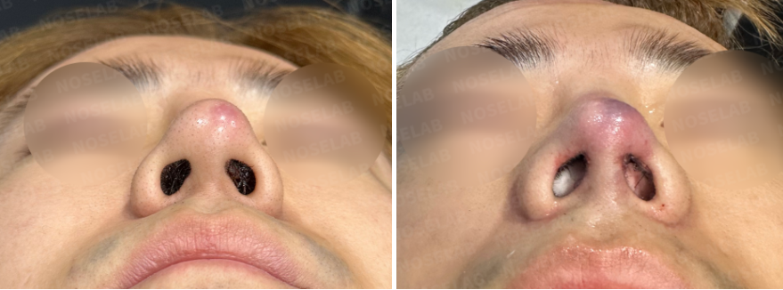
Nostril view: Asymmetry corrected, granulation tissue removed, healthy nostril shape restored.
Immediately after surgery, swelling was reduced and the nasal shape appeared balanced and natural.
Why Autologous Rib Cartilage is Essential in Silicone Exposure Revision
Long-term silicone implants, especially L-shaped, often lead to complications such as extrusion and inflammation. To ensure stable reconstruction, autologous rib cartilage was used for septal extension and alar correction, minimizing the risk of recurrent infection and contracture.


Conclusion
This case demonstrates the importance of immediate treatment in patients with silicone exposure rhinoplasty complications. Complete implant removal, infection control, and rib cartilage reconstruction were key to restoring both function and aesthetics.
At Nose Lab Clinic, we specialize in closed rhinoplasty and complex revision cases, ensuring safe and natural outcomes with autologous tissue.
This has been Dr. Cha-Young Kang, head surgeon at Nose Lab Clinic.
Thank you.

Messenger(WhatsApp) : +82 1057360302
Home page : www.noselab.co.kr
Instagram : noselab_global
YouTube : Noselab
Email : noselab@naver.com


댓글